Introduction
Lichens, also known as lichenized fungi, are an iconic example of symbiosis and belong to Ascomycota Caval.-Sm. or Basidiomycota R. T. Moore (Lücking, Hodkinson, & Leavitt,2017). Their thalli consist mainly of the hyphae of the lichenized fungus (called mycobiont in symbiosis) as well as various heterotrophic and autotrophic eukaryotic and prokaryotic organisms. The mycobiont is associated with one or more photosynthetic partners (photobionts), which may belong to eukaryotic green algae or prokaryotic cyanobacteria (e.g., Dal Grande et al., 2017; Friedl & Büdel, 2008; Lücking et al., 2009; Moya et al., 2017; Onuț-Brännström et al., 2018; Purvis, 2000). Lichen thalli also harbor other fungi, and this mycobiome is formed by the following three groups: (i) symptomatic lichenicolous fungi (i.e., showing their reproductive structures, which distinguishes them from endolichenic fungi), (ii) asymptomatic endolichenic fungi, and (iii) extraneous fungi and their diaspores (e.g., Arnold et al., 2009; Bates et al., 2012; Diederich et al., 2018; Fernández-Mendoza et al., 2017; Flakus et al., 2019; Muggia et al., 2016; Spribille et al., 2016). The third group probably does not play any ecological role in lichen symbiosis; however, these lichen thalli may serve as banks of spores and mycelia or reservoirs of the local fungal mycobiome (Fernández-Mendoza et al., 2017). Lichen thalli also contain nonphotosynthetic bacteria, which contribute to the functioning of the symbiotic system of the lichen as a whole (e.g., Bates et al., 2011, 2012; Grube et al., 2015; West et al., 2017).
The most common lichen photobionts belong to Chlorophyta Reichenbach, and nearly 90% of lichenized fungi are associated with green algae (chlorobionts), mostly of the genera Asterochloris Tschermak-Woess, Trebouxia Puymaly, and Trentepholia Mart. s. l. (e.g., Dal Grande et al., 2017; Friedl & Büdel, 2008; Kosecka et al., 2020; Miadlikowska et al., 2006, 2018; Moya et al., 2017; Onuț-Brännström et al., 2018; Rivas Plata et al., 2010; Singh et al., 2019). Some lichenized fungi form thalli with other autotrophic groups of organisms, of which cyanobacteria (cyanobionts) are the commonest symbiotic partners (e.g., Lücking et al., 2009, 2017; Miadlikowska et al., 2006, 2018; Tschermak-Woess, 1988, 1989). In some lichens, both types of photobionts can be present in a single thallus, forming a tripartite association, with green algae being the dominant component of the thallus and cyanobionts recruited in cephalodia (e.g., Lamb, 1951; Miadlikowska et al., 2018; Oset & Kukwa, 2012; Schneider et al., 2016; Tønsberg & Goward, 2001). In a few genera, a single species of lichenized fungus can form two different thalli (photomorphs), with either a chlorobiont (chloromorph) or a cyanobiont (cyanomorph), and this type of association is related to environmental factors (Green et al., 1993; Moncada, Coca, & Lücking, 2013; Purvis, 2000; Ranft et al., 2018; Tønsberg & Goward, 2001). More recently, it was reported that a single thallus of one lichen can associate with numerous species or operational taxonomic units (OTUs) of closely related photobionts and that one or two strains are dominant within the thallus, with the rest of the pool representing associated photobionts (e.g., del Campo et al., 2013; Dal Grande et al., 2017; Moya et al., 2017; Onuț-Brännström et al., 2018).
In general, the morphology of the lichen thallus is shaped by the mycobiont, but in a few genera of filamentous lichens (e.g., Cystocoleus Thwaites or Cyphellostereum D. A. Reid), the thallus structure depends upon the photobiont filaments and the fungal hyphae surrounding them (e.g., Dal-Forno et al., 2013; Hawksworth et al., 2011). However, in some lichen groups with nonfilamentous photobionts, the morphology of the thallus depends upon autotrophic partners, but in a different way. In other words, the switch from one type of photobiont to another changes the morphological traits (anatomic structure, color, and size of the thallus) or even propagation modes (asexual versus sexual reproduction). Sometimes, in such cases, two names were applied to different morphotypes of the same species (e.g., Ertz et al., 2018; Heidmarsson et al., 1997; Jørgensen, 1998; Miadlikowska et al., 2018; Moncada, Coca, & Lücking, 2013; Tønsberg & Goward, 2001). Nevertheless, the taxonomy and systematics of lichens are based always, according to Article F.1.1. of the International Code of Nomenclature for Algae, Fungi, and Plants, on the fungal partner, and only one name can be applied (Turland et al., 2018). In this review, some examples of the influence of shift in photosynthetic partners on the taxonomy of lichenized fungi are presented.
Sticta – A Case of Fishy Lichens
Sticta (Schreb.) Ach. is a genus of large, foliose (rarely fruticose) lichens developing cyphellae (structures on the lower side of thalli that allow gas exchange) – known only in this genus (Moncada, Lücking, & Betancourt-Macuase, 2013; Purvis, 2000). The genus is widely distributed but is more diverse in the tropics and southern hemisphere (e.g., Galloway, 1994, 1997; Lücking, Hodkinson, & Leavitt, 2017; Moncada, Lücking, & Betancourt-Macuase, 2013; Ranft et al., 2018). Similar to many members of Peltigeraceae Dumort., Sticta species can associate either with green algae or cyanobacteria. Moreover, similar to that in Peltigera Willd. or Ricasolia De Not., in some Sticta species, the cyanobiont is enclosed in cephalodia or two different photomorphs (chloromorph and cyanomorph; Figure 1A) develop (e.g., Galloway, 1994, 1997, 1998, 2001; Miadlikowska et al., 2018; Moncada, Coca, & Lücking, 2013; Moncada, Lücking, & Betancourt-Macuase, 2013; Ranft et al., 2018; Tønsberg & Goward, 2001). Thalli with cyanobionts also have a very characteristic fish-like smell caused by the production of soluble amines (Galloway & Nash, 2004; James & Henssen, 1976).
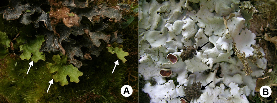
Figure 1
(A) Two photomorphs of Stica on the same thallus with chloromorph (arrow) growing out of cyanomorph. (B) Dendriscocaulon-like cephalodia (arrow) on thallus of Ricasolia amplissima in Scotland.
The taxonomy of Sticta is based on numerous morphological characteristics (e.g., type of thallus, color of the lower surface, and development of the tomentum); however, the type of photobiont was typically the first characteristic in keys dividing species into groups (Galloway, 1994, 1997, 1998). This morphology-based taxonomy has obscured the diversity and identity of species, with photomorphs recognized as separate taxa and some of them even placed in different genus, Dendriscocaulon Nyl. (e.g., Galloway, 2001; Magain et al., 2012; Moncada, Coca, & Lücking, 2013; Purvis, 2000; Ranft et al., 2018; Tønsberg & Goward, 2001; see also below).
One of the first discoveries that the same Sticta species can recruit either a green alga or a cyanobacterium was based on morphological and ecological studies. It was found that environmental factors (light and humidity) are crucial to the selection of photobiont type for thallus development. In more humid and sheltered habitats with low illumination, a cyanobiont is preferred over a chlorobiont, which is the photosynthetic component of the thallus in more dry and open habitats. In intermediate conditions, composite thalli are found, i.e., thalli with chlorobionts growing on thalli with cyanobionts (James & Henssen, 1976). These observations were later confirmed by laboratory experiments and molecular data, and several additional pairs of cyanomorphs and chloromorphs were detected and described (e.g., Armaleo & Clerc, 1991; Galloway, 2001; James & Henssen, 1976; Magain et al., 2012; Moncada, Coca, & Lücking, 2013; Purvis, 2000; Ranft et al., 2018; Stocker-Wörgötter, 2002). Moreover, different photobionts can be recruited when the environment changes due to natural disasters, e.g., falling of trees to form open habitats. At a few sites in Bolivia, thalli with cyanobacteria were deteriorating and being overgrown by the attached thalli with green algae (Kukwa, unpublished; Figure 1A).
Shift in photobiont type may alter the morphology, such as color of the thallus or tomentum (Moncada, Coca, & Lücking, 2013) as well as thallus organization. Some species, when associated with cyanobacteria, form fruticose and branched thalli, which have been placed in the genus Dendriscocaulon (Galloway, 2001; James & Henssen, 1976; Magain et al., 2012; Purvis, 2000; Ranft et al., 2018; Tønsberg & Goward, 2001). Interestingly, however, Dendriscocaulon-like cyanomorphs of the genus Sticta are mostly found in temperate regions, whereas thalli with cyanobacteria are foliose and more similar in morphology to their green algal counterparts in the tropics (Moncada, Coca, & Lücking, 2013).
In a very few cases (e.g., Sticta phyllidiokunthii Moncada & Lücking), the switch of photobionts also alters the mode of reproduction, as cyanomorphs develop vegetative propagules in addition to apothecia, whereas chloromorphs develop apothecia alone (James & Henssen, 1976; Moncada, Coca, & Lücking, 2013).
Dendriscocaulon – A Heterogenic Assemblage of Species
Dendriscocaulon, introduced by Nylander (1885) for D. bolacinum (Ach.) Nyl., accommodates species with branched thalli and cyanobionts. Numerous species have been described, but several studies have also reported unnamed Dendriscocaulon-like thalli (often as cephalodia) associated with lichens containing green algae as the main photobiont (e.g., Clerc et al., 1992; Galloway, 1983, 2001; James & Henssen, 1976; Magain et al., 2012; Tønsberg & Goward, 2001). The association between both photomorphs was speculated for years, although in some cases, it was difficult to prove that they represented the same species when cyanomorph and chloromorph were allopatric (James & Henssen, 1976). Thanks to molecular techniques, this problem could be solved. Sequencing of fungal molecular markers showed that Dendriscocaulon-like lichens are, in fact, cyanomorphs of species accommodated in the genera Dendriscosticta Moncada & Lücking, Sticta, and Ricasolia (Figure 1B) and that the genus itself is a heterogeneous assemblage of distantly related species (e.g., Galloway, 2001; Magain et al., 2012; Moncada, Coca, & Lücking, 2013; Moncada, Lücking, & Betancourt-Macuase, 2013; Ranft et al., 2018; Stenroos et al., 2003). Therefore, formally described Dendriscocaulon species have now become synonyms of other species and the generic name itself is a synonym of Ricasolia (Galloway, 2001; Moncada, Lücking, & Betancourt-Macuase, 2013).
Symbiosis with two different photobionts may also have chemical consequences. James and Henssen (1976) studied 100 free-living Dendriscocaulon-like thalli using thin-layer chromatography or microcrystal tests and found no lichen secondary metabolites. However, the chloromorph may produce a few lichen substances, e.g., scrobiculin present in the chloromorphs of Ricasolia amplissima (Scop.) De Not. s. l. (for the most recent taxonomy of the species, see Cornejo et al., 2017). The nature of this phenomenon is unknown, but perhaps, the presence of cyanobacteria actively inhibits the production of certain lichen substances (James & Henssen, 1976). Meanwhile, this type of photobiont also alters the products of nitrogen metabolism: Ammonia and amines are produced by cyanomorphs and, when the thallus is wet, are released, emitting fish-like odor (James & Henssen, 1976; see also previous section).
Buellia violacefusca and Lecanographa amylacea – When Two Become One
A single lichen thallus of the same species can contain numerous species or OTUs of closely related photobionts, and different thalli may also be associated with different phylogenetic lineages of photobionts of the same genus (e.g., del Campo et al., 2013; Dal Grande et al., 2017; Friedl, 1987; Guzow-Krzemińska, 2006; Moya et al., 2017; Muggia et al., 2014; Nelsen & Gargas, 2008; Onuț-Brännström et al., 2018). Occasionally, although a lichen recruits photobionts of two closely related green algal genera (Engelen et al., 2010), there are no changes in morphology, reproductive methods, or secondary metabolites. However, the shift in the symbiont from the green algal genus Trebouxia in the class Trebouxiophyceae Friedl to Trentepohlia s. l. in the class Ulvophyceae K. R. Mattox & K. D. Stewart may alter the reproductive mode and phenotypic dimorphism (Ertz et al., 2018).
Buellia violaceofusca G. Thor & Muhr was described as a sterile, sorediate lichen with blue-brown soralia and green algae as the photobiont. Due to the absence of apothecia, its taxonomic position was unclear, but owing to morphological similarity to Buellia griseovirens (Turner & Borrer ex Sm.), it was tentatively placed in the genus Buellia De Not. (Thor & Muhr, 1991) in the class Lecanoromycetes O. E. Erikss. & Winka (Wijayawardene et al., 2020). When fresh material of the species became available, it was sequenced. Surprisingly, analyses of fungal mtSSU and ITS rDNA markers placed it within the genus Lecanographa Egea & Torrente in the class Arthoniomycetes O. E. Erikss. & Winka (Ertz et al., 2018). Sequences appeared to be identical to Lecanographa amylacea (Ehrh. ex Pers.) Egea & Torrente, which lacks soredia, but reproduces sexually, forming thalli with a trentepohlioid photobiont that contains large amounts of carotenoid pigments, causing the algae to appear yellow-orange (Ertz et al., 2018). Buellia violaceofusca and Lecanographa amylacea are conspecific, the latter being the oldest available name for this lichenized fungus (Ertz et al., 2018).
This is a well-documented example of a lichen species recruiting two different green algae of distantly related genera. A switch between these unrelated photosynthetic partners is obviously responsible for thallus dimorphism and has a major impact on the anatomy, morphology, and reproductive strategy of the species. The Trebouxia-morphotype of Lecanographa amylacea is always sterile and sorediate, contrary to its trentepohlioid morphotype, which never produces soredia. When Lecanographa amylacea is fertile, its ascospores can capture Trebouxia algae from other lichens (or perhaps also from free-living, nonlichenized Trebouxia) to form the sorediate thallus, but when trentepohlioid algae are recruited, it develops esorediate, fertile thalli. This flexibility can be considered as a strategy to increase habitat tolerance, which allows the lichen to withstand environmental changes. Examination of numerous specimens revealed that the sorediate component developed when the esorediate morphotype of Lecanographa amylacea grew in proximity to Chrysothrix candelaris (L.) J. R. Laundon; soredia developed in the areas where hyphae of the former invaded the thallus of the latter. Molecular data showed that both species often share the same Trebouxia strain (Ertz et al., 2018).
Lecanographa amylacea is a rare lichen, but its sorediate morphotype is more common and reported from stands where fertile thalli do not grow (e.g., Ertz et al., 2018; Kukwa et al., 2012; Poelt, 1994; Thor & Muhr, 1991). This observation indicates that the strategy to recruit two different photobionts and the shift of the reproductive mode can be evolutionarily advantageous.
This is the first and thus far the only documented case of a single lichen species recruiting Trebouxia and Trentepohlia s. l. photobionts, which have altered its morphology and reproductive mode. However, by studying sterile sorediate or isidiate lichens of the family Graphidaceae in Bolivia, we found that this phenomenon may exist in a few cases within this group of lichenized fungi. Most members of Graphidaceae form thalli with trentepohlioid photobionts (e.g., Kosecka et al., 2020; Rivas Plata et al., 2010; Staiger, 2002), but in some sterile Bolivian samples of this group, Trebouxiophyceae photobionts were detected using morphological and molecular approaches. However, their fertile photomorph with Trentepholia-like algae have not been found yet. More details will be presented in a forthcoming article.
Ionaspis/Hymenelia complex – Is the Photobiont Not Enough to Distinguish Genera?
The genera Ionaspis Th. Fr. and Hymenelia Kremp. (Hymeneliaceae Körb.) were traditionally separated on the basis of their different photobionts, trentepohlioid or trebouxioid, respectively. However, Eigler (1969) showed heterogeneity within this complex, with some Ionaspis species being more similar to Hymenelia species than to their congeners. Therefore, the distinction of these genera based on the photobiont was questioned by numerous lichenologists (e.g., Eigler, 1969; Jørgensen, 1989; Lutzoni & Brodo, 1995). By employing cladistic analyses of morphological, anatomical, and allozyme data, Lutzoni and Brodo (1995) reviewed North American taxa within the Ionaspis/Hymenelia complex and reclassified some species. They treated the photobiont as a minor character and separated these two genera on the basis of differences in epihymenial pigments, ascospore width, and hymenium thickness (Lutzoni & Brodo, 1995). Although their new classification is generally accepted, the distinction between these two genera was claimed to be practically difficult and artificial compared to the photobiont-based classification, and the need for morphological and molecular revisions of these taxa was thus highlighted (Fryday & McCarthy, 2018; Kantvilas, 2014). However, this complex has not been studied in more detail using DNA data, with the exception of a study based on limited sampling from Alaska, which showed that this group of lichens warrants further investigation (McCune et al., 2018).
Stictidaceae and Optional Lichenization
The family Stictidaceae Fr. comprises 28 genera (Wijayawardene et al., 2020) of lichenized, lichenicolous, and saprotrophic fungi (e.g., Diederich et al., 2018; Wedin et al., 2004, 2005, 2006). This also includes the genus Stictis Pers., which originally comprised epixylic, nonlichenized saprotrophic fungi (Wedin et al., 2004, 2005, 2006). However, molecular data also placed species of the former Conotrema Tuck., which are lichenized and epiphytic fungi, within the genus Stictis (Wedin et al., 2004, 2005, 2006). Although such a situation is not an exception in fungi, it is remarkable in this case. Using molecular markers, Wedin et al. (2004, 2005, 2006) discovered that neither the genera nor the species are monophyletic. Moreover, the same species, depending on the substrate (bark or wood), either formed a lichenized Conotrema-like thallus or lived as a saprotroph without symbiotic algae (Stictis-like thallus) and completed its lifecycle. The phenomenon that a single species can exist as a lichen or a saprotroph is called optional lichenization (Wedin et al., 2004, 2005, 2006).
Individuals of Stictis species that can form lichenized and nonlichenized thalli differ in their morphology. Ascomata of both forms are very similar, but as saprotrophs, they have generally more heavily pigmented ascomatal walls and are more exposed on the substrate. Additionally, individuals living in symbiosis with algae form highly visible, whitish, lichenized thalli on tree barks, whereas thalli of individuals lacking photobionts are less evident, completely immersed in the substratum and only their ascomata are visible. A very similar situation, with two nutritional modes (lichenized and saprotrophic), is also known in another member of Stictidaceae: Schizoxylon albescens Gilenstam, H. Döring & Wedin (Fernández-Brime et al., 2019; Wedin et al., 2006).
Optional lichenization may play important roles in the evolution of Stictidaceae, representing an advantageous adaptive strategy allowing the species to grow on different substrates. It enables the species to colonize tree barks (mostly Populus tremula) when there is no lignum available for the nonlichenized form in the habitat or when the lignum is already present but the host trees are too young for colonization of the lichenized form (Wedin et al., 2004). Moreover, species with two different nutritional modes may be more common and widespread than species lacking this potential (Wedin et al., 2004, 2006).
Conclusions
The switch of photobionts or optional lichenization are the two potential evolutionarily advantageous strategies that allow lichens to occupy a wider range of habitats. Recruitment of different photobionts or optional lichenization allows the species to colonize habitats with diverse conditions of light and humidity (e.g., Ertz et al., 2018; Green et al., 1993; James & Henssen, 1976; Purvis, 2000) or occupy different substrates (Fernández-Brime et al., 2019; Wedin et al., 2004, 2005, 2006). Both these strategies alter the anatomy, morphology, secondary metabolites, and reproductive modes (Ertz et al., 2018; James & Henssen, 1976; Lutzoni & Brodo, 1995; Magain et al., 2012; Moncada, Coca, & Lücking, 2013; Purvis, 2000; Wedin et al., 2004) as well as influence the taxonomy and classification of lichenized fungi.
According to Turland et al. (2018), the taxonomy of lichens is always based on the fungal partner (Article F.1.1.); therefore, only a single name must be used for a fungus associated with different photobionts (or in the case of optional lichenization, lacking photobionts), despite variations in the morphology, chemical composition, and reproductive modes caused by association with different photobionts. However, before the application of molecular methods, the identification and classification of such different morphs of a single species were limited and often separate names were applied (e.g., Ertz et al., 2018; Heidmarsson et al., 1997; Jørgensen, 1998; Laundon, 1995; Tønsberg & Goward, 2001). Laundon (1995) proposed forma as a taxonomic rank to recognize photomorphs; however, Heidmarsson et al. (1997) claimed that it was illogical to use any taxonomic rank in such cases, particularly when two photomorphs can form an association in a single lichen thallus [like in Sticta canariensis (Bory) Bory ex Delise]. These authors also stated that any rank should reflect genetic differences – a point of view accepted in subsequent studies (e.g., Ertz et al., 2018; Miadlikowska et al., 2018; Moncada, Coca, & Lücking, 2013; Purvis, 2000; Ranft et al., 2018).
Handling Editor
Agnieszka Popiela, University of Szczecin, Poland; https://orcid.org/0000-0001-9297-0538
Authors’ Contributions
MKu prepared the core parts of introduction, first second, third, and sixth chapters, and conclusions; MKu, MKo, and BGK prepared the fourth chapter; BGK prepared fifth chapter; MKo provided data on the photobionts of Graphidaceae; all authors read and corrected the entire text
