. Introduction
Medicinal plants have played a crucial function in conventional medicine for a long time, offering a wide range of therapeutic advantages due to their many bioactive components (Chaachouay & Zidane, 2024). The plants contain a diverse array of phytochemicals, such as flavonoids, alkaloids, and glycosides, which are accountable for their diverse medicinal properties (Agidew, 2022). Medicinal plants hold significant importance not only in history but also in contemporary research, which validates and expands our understanding of their potential to address various health issues (Jacob et al., 2024).
In modern medicine, the investigation of medicinal plants has resulted in notable breakthroughs in the treatment of chronic diseases, enhancement of general well-being, and prevention of disorders (Yuan et al., 2023). The use of medicinal plants in modern treatment methods exemplifies the ongoing partnership between conventional knowledge and scientific advancement (David et al., 2015; Chao et al., 2017). Nonetheless, the growing demand for natural health products has highlighted the importance of medicinal plant conservation and appropriate use (Sen & Samanta, 2015; Van Wyk & Prinsloo, 2018).
Studies emphasized the antioxidant, anti-inflammatory, antibacterial, and neuroprotective characteristics of these plants (Mucha et al., 2021; Vaou et al., 2021; Rehman et al., 2019). Plant-derived bioactive compounds have antioxidant qualities that enable them to mitigate conditions such as cardiovascular illnesses, neurological disorders, and infections (Akbari et al., 2022; El-Saadony et al., 2023; Samtiya et al., 2021). This is due to their capacity to lessen oxidative stress and counteract harmful reactive oxygen species (ROS) (Adwas et al., 2019). Antioxidants are very important for protecting cells and maintaining cellular balance. Various antioxidants derived from plants help to prevent oxidative damage to DNA, lipids, and proteins, which contribute to the development of various diseases (Adwas et al., 2019; Rahal et al., 2014; Kurutas, 2015).
Certain chemicals found in plants, like polyphenols and flavonoids, are very good at protecting cells from oxidative damage. They do this by fighting reactive oxygen species (ROS) and other harmful molecules. Among the classes of plant-based compounds that have the greatest promise for future scientific advancements are polyphenols and flavonoids. They are plant chemicals that function as potent antioxidants, effectively protecting cellular structures from oxidative damage and counteracting reactive oxygen species (ROS) (Pandey & Rizvi, 2009). Numerous studies (Jafernik et al., 2024; de Lima Cherubim et al., 2020; Srinivasan et al., 2002) have demonstrated their extensive use in makeup and medicine products. Numerous studies (Akbari et al., 2022; El-Saadony et al., 2023; Samtiya et al., 2021) have confirmed the efficacy of these chemical compounds in treating cardiovascular diseases, cancer, infections, and neurological disorders. Polyphenols are the most common secondary molecules in plants. Tannins, flavonoids, proanthocyanidins, phenolic acids, and flavonoids are some of these compounds (Tsimogiannis & Oreopoulou, 2019; Singla et al., 2019).
About thirty (30) tree species make up the genus Boswellia, which is a member of the family Burseraceae. These trees are mostly indigenous to the arid and semi-arid areas of northern Africa, the Arabian Peninsula, and parts of India (Birhan et al., 2023). The most well-known product of these trees is frankincense (olibanum), a fragrant resin that has been utilized for centuries in perfumery, religious ceremonies, and traditional medicine. Boswellia serrata, Boswellia sacra, Boswellia carteri, and Boswellia papyrifera are some of the most researched species; they all produce resin with somewhat distinct pharmacological profiles and chemical compositions (Khalifa et al., 2023; Feng et al., 2021; Wang et al., 2020; Al-Dahmash et al., 2021).
Traditional ethnomedicine applies Boswellia resin topically to treat a variety of skin conditions. Boswellic acids, pentacyclic triterpenoids that are abundant in Boswellia resins, are phytochemicals that have been demonstrated to have various biological actions, such as anti-inflammatory, anti-arthritic, anti-cancer, and antibacterial properties (Feng et al., 2021; Wang et al., 2020). Traditional medicine, including Ayurveda and traditional Arabic medicine, extensively utilizes extracts from Boswellia species due to these properties. They have also drawn interest in contemporary pharmacological research.
Boswellia dalzielii (B. dalzielii) Hutch (Burseraceae) is one of the plant species found in Northern Nigeria and the West African savannah used by traditional healers to treat several disease conditions, including mental illness and skin disorders (Alemika et al., 2006). All parts of B. dalzielii were employed for medical purposes. Traditional medicine in Northern Nigeria uses the bark in large quantities as a wash for fever and rheumatism, and administers the fluid for gastrointestinal difficulties and convulsions, among other uses. Additionally, Dalziel (20061937) noted that fresh bark can alleviate dizziness and palpitations and serves as a tonic. The North-Central region of Nigeria also makes use of the preparation of the stem bark to treat mental disorders (Ibrahim et al., 2007). Previous research has documented the antibacterial, antiulcer, antispasmodic, hepatoprotective, and hypoglycemic activities (Adelakun et al., 2001; Nwinyi et al., 2004; Hassan et al., 2009; Odeghe et al., 2012; Balogun et al., 2013) and behavioural effects (Nazifi et al., 2017) of B. dalzielii stem bark extract.
Recent investigations into B. dalzielii have highlighted its potential as a source of biologically active compounds. Salisu et al. (2017) carried out a comprehensive phytochemical screening of the aqueous stem bark extract using HPLC, FTIR, and GC-MS techniques. Their study identified several functional groups and major chemical constituents such as n-hexadecanoic acid, stearic acid, and 9-hexadecenoic acid, along with significant antimicrobial activity against Staphylococcus aureus, Salmonella typhi, and Pseudomonas aeruginosa, among others. These findings underscore the presence of both polar and non-polar bioactive compounds in the stem bark, which supports the rationale for further phytochemical investigation of this species using more advanced profiling techniques.
Surprisingly, despite numerous traditional uses in ethnomedicine, a detailed comparison of the antioxidant properties of the plant’s leaf, stem bark, and root bark and phytochemical profile study of different parts of B. dalzielii have never been made to find biologically active compounds that would explain the medicinal properties attributed to raw plant material. Therefore, the present study aimed to evaluate the antioxidant activities and comprehensive phytochemical analysis of aqueous extracts from the leaf, stem bark, and root bark of B. dalzielii using various in vitro assays and UHPLC-DAD-MS analysis, providing valuable insights into its therapeutic usage and promoting further research into its medicinal properties. To the best of our knowledge, this study represents the first UHPLC-DAD-MS analysis of Boswellia dalzielii collected from the tropical savanna region of Gadam, northeastern Nigeria.
. Materials and methods
. Chemicals and reagents
To distill water for this investigation, a glass distillation procedure was used. The chromatographic-quality acetonitrile and formic acid were purchased from Merck, which is in Darmstadt, Germany. In preparation for HPLC, water was purified using Millipore Simplicity System gotten from Bedford, Massachusetts, United States of America. We obtained high-grade methanol from POCh, located in Gliwice, Poland. Sigma-Aldrich (St. Louis, MO, USA) provided quercetin 3-O-glucoside (isoquercitrin) (catalog number: 00140585), kaempferol 3-O-glucoside (astragalin) (catalog number: 79851), gallic acid (3,4,5-trihydroxybenzoic acid) (catalog number: 91215), 5-O-caffeoylquinic acid (chlorogenic acid) (catalog number: 00500590), ellagic acid (catalog number: 14668), quercetin 3-O-galloylhexoside (catalog number: 56316-75-7), while Carl Roth (Karlsruhe, Germany) provided isorhamnetin 3-O-glucoside (methoxy quercetin hexoside) (catalog number: Art.9386.1).
. Sample collection
Leaves, stem bark, and root of Boswellia dalzielii (Hutch) were collected in December 2023 from Gadam, Kwami Local Government Area, Gombe State, northeastern Nigeria (11°6'6" E; 10.47484 N, 11.10177 E; Aw: Tropical Savanna climate). The plant samples were authenticated in January 2024 by a qualified taxonomist at the herbarium section of the Department of Plant Science and Biotechnology, Ekiti State University, Ado Ekiti, Nigeria. Voucher specimens were deposited under the herbarium numbers UHAE 2024001a (leaves), UHAE 2024001b (stem bark), and UHAE 2024001c (root bark) for future reference.
. Sample preparation
Fresh leaves, stembark and rootbark of B. dalzielli were cleaned and air dried until constant weights were obtained. Although the initial mass and moisture content of the fresh plant materials was not recorded at the time of sample preparation. The dried stembark and rootbark were reduced to coarse powder using pestle and mortar, then pulverized using electric grinder while the dried leaves were homogenized using a blender (Vitamix 5200, 2-peak horsepower motor). The plant samples were kept separately in an air-tight container prior to the extraction.
. Preparation of aqueous extract
Separately, the ground plant samples (200 g each) were soaked in distilled water (4000 mL) for 24 hours on a rotatory shaker set at 150 rpm. The products were filtered and then freeze-dried. It gave a yield of 15%, 15% and 8.4% (w/w) for leaf, stembark and rootbark respectively. 10 mg of the freeze-dried extract were dissolved in 1 mL of water/methanol (1:1) solution to prepare the crude extract needed for UHPLC-DAD-MS characterization. The next step was to filter the solution using a Chromafil syringe membrane with a thickness of 0.45 µm, which was made in Duren, Germany, and then transfer it to autosampler vials. After that, we kept the vials in a dark place at a temperature of four degrees Celsius. The solution was later injected into the HPLC system and subjected the filtrate to rotary evaporation. For the determination of in vitro antioxidant parameters, 10 mg of the freeze-dried extracts were reconstituted in 1 mL of distilled water.
. UHPLC-DAD-MSn analysis
Dionex UHPLC-3000 RS equipment from Leipzig, Germany, was used to analyze freeze dried water-based extracts of B. dalzielli’s leaves, stembark, and rootbark dissolved in methanol (10 mg/ml). The system was equipped with DAD detector directly linked an AmaZon SL ion trap mass spectrometer with an ESI interface (Bruker Daltonics, Bremen, Germany). All UV-Vis spectra were recorded from 190 to 450 nm. The ESI source of the mass spectrometer had the following conditions 40-psi nebulizer pressure, 9 L/min drying gas flow rate, 134°C nitrogen gas temperature, and a 4.5 kV capillary voltage. By scanning a range of mass-to-charge ratios (m/z) from 70 to 2200, mass spectra were acquired. For the analysis, a chromatography column from Phenomenex in Torrance, California, called Kinetex XB-C18 was used (150 × 2.1 × 1.7 µm). The mobile phases were 0.1% formic acid water (A) and 0.1% formic acid acetonitrile (B), and elution was conducted with the following gradient elution: 0 min, 0% B; 60 min, 26% B; and 70 min, 95% B. The flow rate was 0.3 mL/min, and the injection volume was 3 µL of the prepared extract. The column oven temperature was maintained at 25°C. Compounds were characterized based on the maxima observed in their UV-vis spectra and on their MS spectra. The identification was based on comparison with literature data and with chemical standards.
. Measurement of total phenolic content
Singleton et al. (1999) methodology was used to measure the total phenolic content. The extracts were diluted appropriately and combined with 106.38 µL of a 10% solution of Folin-Ciocalteau’s reagent (volume/volume). The mixture was then neutralized with 85.10 µL of a 7.5% solution of sodium carbonate in a 96-well plate. The process was repeated three times to ensure accuracy. The reaction mixture was incubated for 40 minutes at a temperature of 45°C, and absorbance of the mixture measured at 765 nm with a BioTek Synergy microplate reader (5301 Stevens Creek Blvd., Santa Clara, CA 95051, USA). The total phenolic content was subsequently assessed using gallic acid as the reference standard.
. Measurement of total flavonoid content
Total flavonoid concentrations in the extracts were measured using a modified procedure based on Meda et al. (2005). 40 µL sample was combined with 40 µL of methanol, 4.0 µL of 10% AlCl3, 4.0 µL of 1 M potassium acetate, and 112 µL of water on a 96-well plate. The mixture was diluted as necessary and replicated three times. After incubating at room temperature for 30 minutes, absorbance of the reaction mixture was measured at 415 nm using a BioTek Synergy microplate reader (5301 Stevens Creek Blvd., Santa Clara, CA 95051, USA). The total flavonoid content was subsequently assessed using quercetin as the reference standard.
. Measurement of vitamin C content
According to Benderitter et al. (1998), the process for determining the extracts’ vitamin C concentration comprised multiple steps. Following a thorough mixing, 15.30 µL of a solution containing 230 mg of thiourea, 270 mg of CuSO4•5H2O, 100 mL of 5 M H2SO4, and 2 g of dinitrophenyl hydrazine was added. 40 µL of a properly diluted extract, 20.40 µL of 13.3% trichloroacetic acid (TCA), and 41.90 µL of water made up the reaction mixture. Three replicates of this experiment were conducted on 96-well plates. Following three hours of incubation at 37°C, 0.5 mL of 65% (v/v) Sulfuric acid (H2SO4) was added to the solutions. A BioTek microplate reader (5301 Stevens Creek Blvd., Santa Clara, CA 95051) was used to measure absorbance at 520 nm. Using ascorbic acid as a reference standard, the vitamin C content was determined and represented in milligrams of ascorbic acid equivalent per gram.
. The potential to scavenge DPPH-free radicals
Gyamfi et al. (1999) methodology was used to measure 1,1-diphenyl-2 picrylhydrazyl (DPPH) radical scavenging ability of the extracts. Briefly, the experiment was performed in triplicate on a 96-well plate containing 100 μL of a DPPH solution (0.4 mM in methanol) with various doses of extracts or vitamin C (dissolved in distilled water) (0, 0.83, 1.67, 2.50, 3.33, and 4.17 mg/mL). Absorbance of the reaction mixture was measured at 516 nm after 30 minutes of incubation in the dark. The ability of the extract to scavenge DPPH free radicals (in %) was calculated using the following mathematical expression:
. Oxidation of deoxyribose using Fenton’s reaction
Hydroxyl (OH*) free radical scavenging capacity of the extracts was evaluated using the Halliwell and Gutteridge (1981) method. In a reaction mixture comprising 69.58 µL of distilled water, 10.43 µL of 20 mM deoxyribose, 34.78 µL of 0.1 mM phosphate buffer, 3.48 µL of 20 mM hydrogen peroxide, and 3.48 µL of freshly prepared iron (II) tetraoxosulphate (VI), freshly prepared extracts of B. dalzielli were added at concentrations of 0, 1.0, 2.0, 3.0, and 4.0 mg/mL. In this experiment, which was carried out in three replications on a 96-well plate, the reaction mixture was incubated for thirty minutes at 37°C. After that, 43.47 µL of a 2.8% trichloroacetic acid solution were added to halt the reaction. After adding 34.78 µL of a 0.6% TBA solution, the plate was incubated for 20 minutes at 100°C. Equation 1 was utilized to calculate the extracts’ capacity to neutralize hydroxyl radicals, and the absorbance at 532 nm was measured to confirm the results.
. Nitric oxide scavenging assay
The Igbinosa et al. (2011) method was used to assess the extracts’ ability to scavenge nitric oxide (NO*) radical. To (0, 1.0, 2.0, 3.0 and 4.0 mg/mL) of the extract, 100 µL of 25 mM sodium nitroprusside was added in a 96-well-plate in three replicates. The resulting solution was then incubated for two hours at 37ºC, then 50 µL of Griess reagent was added to reaction mixture. The absorbance was quantified at 546 nm, and the mathematical expression (Equation 1) was utilized to quantify the extent of inhibition of the extracts on NO* radical.
. The Ferric-Reducing Antioxidant Power (FRAP) Measurement
Ferric-reducing antioxidant power (FRAP) of the extracts was determined by applying the procedure of Oyaizu’s (1986). This assay determined the extracts’ reducing power, or their capacity to convert Fe3+ to Fe2+. Three aliquots of 9.09 µL of the extract or standard, 22.72 µL of 0.2 M phosphate buffer (pH 6.6), and 0.5 mL of a 1% (w/v) potassium ferricyanide solution (K3[Fe(CN)6]) were all included in the reaction mixture. After combining the ingredients, they were incubated for 20 minutes at 50°C. Following that, 22.72 µL of 10% (w/v) trichloroacetic acid was added to stop the reaction. After that, 18.18 µL of a 0.1% (w/v) FeCl3 solution was added to the mixture after it had been diluted with 90.94 µL of distilled water. At 700 nm, the absorbance of the blue complex was measured. As a reference point, distilled water was utilized in place of the sample and standard in a reagent blank that contained all other components. Utilizing vitamin C as the reference, the ferric-reducing antioxidant capacity was determined.
. The Cupric-Reducing Antioxidant Capacity (CUPRAC) Measurement
Using the protocol described by Apak et al. (2008), the extracts’ cupric-reducing antioxidant capacity (CUPRAC) were evaluated. Using a Cu (II)-neocuproine chromogenic reagent, extracts are tested for their capacity to reduce it to a Cu(I)-Neocuproine complex, which has an orange-yellow appearance with a peak absorbance at 450 nm. Using a reaction mixture containing 40 µL of CUPRAC reagent and different concentrations of samples and controls (0.25, 0.5, 1, 2, and 4 mg/mL), the test was carried out on a 96-well plate. A 1:1:1:0.6 mixture of 10 mM CuCl2·2H2O, 7.5 mM Neocuproine in methanol, 0.1 M ammonium acetate buffer (pH 7.0), and distilled water was used to prepare the CUPRAC reagent. For thirty minutes, the mixture was incubated in the dark. The results were compared to a reagent blank that contained every component except the sample or standard, which was substituted with distilled water. Absorbance was measured at 450 nm.
. Measurement of Iron (Fe2+) chelation capacity
Modified method of Minotti and Aust’s (1987) was used to assess the extracts’ ability to chelate Fe2+. 30 µL of freshly prepared 500 µM FeSO4 was added to the reaction mixture, which already contained 33.6 µL of 0.1 M Tris-HCl (pH 7.4) and 43.6 µL of 0.9% saline. There were also several extract concentrations and/or standard (EDTA) (at levels of 0.83, 1.67, 2.50, 3.33, and 4.17 mg/mL) included. This reaction was run in triplicate in a 96-well plate. After fifteen minutes incubation at room temperature, 2.6 microliters of a 1,10-phenanthroline 0.25% (w/v) solution were added to it. The absorbance was read at 510 nm. Equation 1 was then utilized to calculate the Fe (II)-chelating ability.
. Measurement of Copper (Cu2+) chelation capacity
Saiga et al. (2003) procedure was used for assessing the extracts’ capacity to bind Cu2+ ions. In this procedure, 80 µL of a pH 5.6 0.1 M acetate buffer was combined with 80 µL of a 1 mg/mL CuSO4 solution.The experiment was run in triplicate in a 96-well plate with different extract and/or EDTA (standard) concentrations (0, 0.83, 1.67, 2.50, 3.33, and 4.17 mg/mL). Thirty minutes later, forty microliters of a solution containing four millimoles per liter of pyrocatechol were added. Following a 10-minute incubation period, the sample’s absorbance at 632 nm was measured. In contrast, the control sample was made up of 40 µL of pyrocatechol solution at a concentration of 4 mM and 80 µL of 1 mg/mL CuSO4. The blank was made with a pH 5.6 acetate buffer. Equation 1 was then used to calculate the samples’ Cu2+ chelating capability.
. Determination of the scavenging capacity 50 (SC50) values of the extract
Scavenging Capacity 50 (SC50 – extract concentration causing 50% effectiveness) values of the extracts were calculated using non-linear regression with GraphPad Prism version 8.0.2 for windows by fitting dose-response curves to the experimental data. SC50 values were determined from the graphs as the plant extract concentration reducing radical concentration to half of its initial value, i.e., neutralizing 50% of the free radicals present in a test system. A lower SC50 value indicates higher radical scavenging potential.
. Results
. Percent yields, total phenolic, total flavonoid, vitamin C, and ferric-reducing antioxidant properties of the plant’s aqueous extract
Table 1 displays the percentage yields achieved for the plant extracts examined. Table 1 showed that the leaf extract had the maximum extractive value, followed by the stembark extract, while the rootbark extract had the lowest yield. The findings also showed that the water-based extract of different parts of Boswellia dalzielli had different amounts of phenolic compounds, flavonoids, vitamin C, and ferric-reducing antioxidants (Table 1). The analysis revealed that the aqueous extract of every plant part had a significant concentration of total phenolic content, as indicated in Table 1. The rootbark aqueous extract exhibited a total phenolic content of 31.43 mg GAE/g, while the leaf aqueous extract had the highest value at 57.92 mg GAE/g. The stembark has the highest total phenolic content, followed by the leaf and then the rootbark (P < 0.05).
Table 1
Percentage yield, total phenolic content, total flavonoid content, vitamin C content and ferric reducing antioxidant property of aqueous extract of different parts of Boswellia dalzelli.
Furthermore, Table 1 shows an extremely high total flavonoid concentration of the extract, expressed in terms of the quercetin equivalent. The leaf aqueous extract had a significantly higher concentration than the stembark aqueous extract, while the rootbark aqueous extract had the lowest concentration. Additionally, the leaf aqueous extract had the highest concentration of vitamin C, measuring 27.25 mg AAE/g. The stembark aqueous extract, at 20.71 mg AAE/g, and the rootbark aqueous extract, at 7.68 mg AAE/g, followed closely behind. The vitamin C level was highest in the leaf, followed by the stembark, and then the rootbark (P < 0.05). The reducing power of the water extract from different parts of B. dalzelli is shown in Table 1, with values given in terms of ascorbic acid equivalent (AAE). The stembark aqueous extract exhibited the greatest reducing power (P < 0.05), followed by the leaf aqueous extract, but the rootbark aqueous extract demonstrated the lowest reducing power.
When compared to other plant parts, the rootbark aqueous extract consistently showed the lowest total phenolic, flavonoid, and vitamin C contents, as well as the lowest ferric reducing power activity.
. Fe2+ – chelating capacity of an aqueous extract from various sections of Boswellia dalzielli
Figure 1 shows how an aqueous extract from different parts of B. dalzielli can chelate iron (II). Table 2 presents the calculated SC50 values. The extract chelates iron (II) in a concentration-dependent way, as shown in Figure 1. Nevertheless, there was no significant difference (p < 0.05) in the percentage of chelation between the leaf extract at concentrations of 0.83 and 1.67 mg/mL compared to the control (EDTA). The leaf extract had the lowest SC50 value of 1.33 ± 0.01 mg/mL, whereas the stembark extract had a slightly higher SC50 value of 1.90 ± 0.01 mg/mL. The rootbark extract had the lowest iron (II) chelating ability, and its SC50 value was 2.45 ± 0.03 mg/mL, which was much higher than those of the other two plant parts (p < 0.05). EDTA had a significantly lower SC50 value (1.07 ± 0.01 mg/mL) in comparison to plant components (p < 0.05).
Figure 1
(a) Fe2+ – chelating ability of aqueous extract of different parts of Boswellia dalzelli. Results are illustrated as mean ± S.D from 3 measurements. Results are significant at *p < 0.05 compared with EDTA at 0.83 mg/ml; #p < 0.05 compared with EDTA at 1.67 mg/ml; $p < 0.05 compared with EDTA at 2.50 mg/ml; &p < 0.05 compared with EDTA at 3.33 mg/ml; @p < 0.05 compared with EDTA at 4.17 mg/ml.
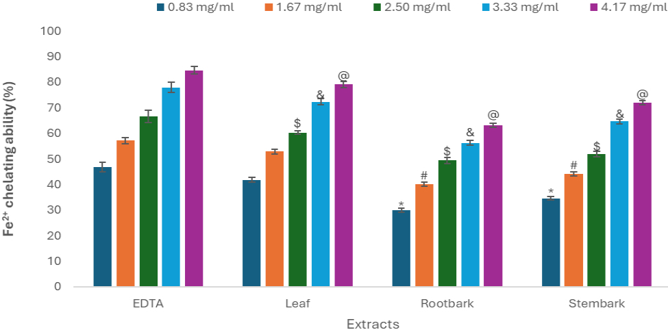
Table 2
Effective concentration causing 50 % scavenging capacity (SC50 values in mg/mL) of 1,1-diphenyl-2 picrylhydrazyl (DPPH), hydroxyl (OH), nitric oxide (NO) radical scavenging abilities, Cupric-reducing antioxidant, copper and iron chelating abilities of aqueous extract of different parts of Boswellia dalzielli.
[i] The radical scavenging abilities of the plant parts were determined as described and expressed as percentage. The SC50 (effective concentration causing 50% scavenging capacity) were calculated using nonlinear regression analysis. Values represent mean ± standard deviation (n = 3). Mean values with the same letter within a row are not significantly different (p > 0.05). Positive control (standard inhibitors): vitamin C and EDTA (Ethylenediaminetetraacetic acid) were used for DPPH*, OH*, NO*, CUPRAC and Cu2+, Fe2+ chelating ability respectively.
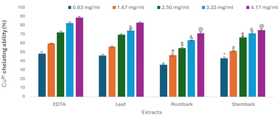
. Cu2+ – chelating capacity of an aqueous extract from various sections of Boswellia dalzielli
Figure 2 demonstrates the ability of an aqueous extract from various sections of B. dalzielli to chelate copper (II). We plotted the percent copper chelating activity of the investigated extracts against their concentration in mg/mL (Figure 2). Table 2 presents the calculated SC50 values. The extract forms a complex with the copper (II) ion in a concentration-dependent way, as illustrated in Figure 2. Nevertheless, the percentage of chelation exhibited by the leaf extract at concentrations of 0.83, 1.67, and 2.50 mg/ml did not show any significant difference (p < 0.05) compared to the control (EDTA). The leaf extract had the lowest SC50 value of 1.08 ± 0.00 mg/ml, whereas the stembark extract had a slightly higher SC50 value of 1.26 ± 0.02 mg/mL. Compared to the other two plant parts, the rootbark extract had the least copper (II) chelating activity, with an SC50 value of 1.79 ± 0.03 mg/mL (p < 0.05). EDTA exhibited a substantially lower SC50 value (0.98 ± 0.01 mg/mL) compared to plant components (p < 0.05), although its SC50 value was not significantly lower than that of the leaf aqueous extract.
Figure 2
Cu2+ – chelating ability of aqueous extract of different parts of Boswellia. Results are illustrated as mean ± S.D from 3 measurements. Results are significant at *p < 0.05 compared with EDTA at 0.83 mg/ml; #p < 0.05 compared with EDTA at 1.67 mg/ml; $p < 0.05 compared with EDTA at 2.50 mg/ml; &p < 0.05 compared with EDTA at 3.33 mg/ml; @p < 0.05 compared with EDTA at 4.17 mg/ml.
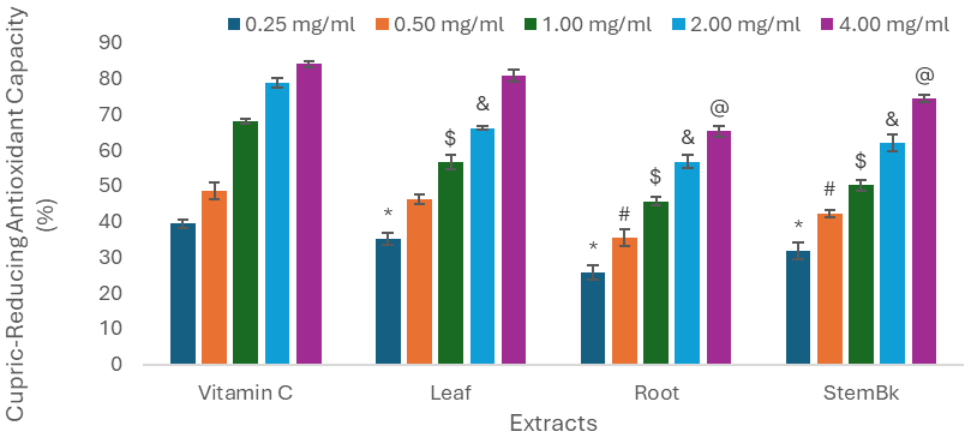
. Cupric-reducing antioxidant capacity of aqueous extract of different parts of Boswellia dalzielli
Table 2 and Figure 3 list the outcomes of the cupric decreasing antioxidant capacity. At a significant level (p < 0.05), the reduction capability for cupric ion of different extracts was found to be in the following ascending order: rootbark < stembark < leaf. When compared to vitamin C, the antioxidant capacity of the plant was found to be much lower in all its parts. However, as the concentration increases, this capacity also increases (Figure 3), indicating that the plant’s parts do have the ability to reduce cupric acid, and that this ability can be increased at higher concentrations. It was discovered that there was no statistically significant difference between the antioxidant potentials of the leaf aqueous extract at 0.50 and 4.00 mg/ml and the control (vitamin C). The rootbark of the plant has the lowest level of antioxidant capacity among its constituent sections. According to the SC50 values (Table 2), the antioxidant capacity of the plant’s components was determined to be highest in the leaf aqueous extract (0.64 ± 0.05 mg/mL) and lowest in the rootbark aqueous extract (1.35 ± 0.13 mg/mL).
Figure 3
Cupric-Reducing Antioxidant Capacity of aqueous extract of different parts of Boswellia. Results are illustrated as mean ± S.D from 3 measurements. Results are significant at *p < 0.05 compared with vitamin C at 0.25 mg/ml; #p < 0.05 compared with vitamin C at 0.50 mg/ml; $p < 0.05 compared with vitamin C at 1.00 mg/ml; &p < 0.05 compared with vitamin C at 2.00 mg/ml; @p < 0.05 compared with vitamin C at 4.00 mg/ml.
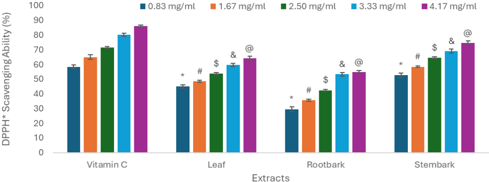
. DPPH* radical-scavenging ability of aqueous extract of different parts of Boswellia dalzielli
Table 2 and Figure 4 present the DPPH* radical-scavenging capacity of the aqueous extract from different sections of B. dalzielli. At a significant level (p < 0.05), the ascending order of the ability of different extracts to scavenge DPPH* radicals was found to be: rootbark < leaf < stembark. All plant portions showed a significant decrease in scavenging ability when compared to vitamin C. However, as the concentration increases, the scavenging capacity also rises (Figure 4), demonstrating the plant’s constituents’ ability to scavenge DPPH* radicals and their amplified capability at higher concentrations. The study observed a substantial statistical difference in the DPPH* scavenging abilities of the plant components compared to the control (vitamin C) at all tested concentrations. Among the constituent sections of Boswellia dalzielli, the rootbark of the plant demonstrates the least DPPH*-scavenging activity. Based on the SC50 values presented in Table 2, the stembark aqueous extract had the highest antioxidant capacity (0.77 ± 0.02 mg/mL), while the rootbark aqueous extract had the lowest antioxidant capacity (3.26 ± 0.01 mg/mL) when compared to vitamin C (0.62 ± 0.01 mg/mL).
Figure 4
DPPH radical-scavenging activity of aqueous extract of different parts of Boswellia dalzelli. Results are illustrated as mean ± S.D from 3 measurements. Results are significant at *p < 0.05 compared with vitamin C at 0.83 mg/ml; #p < 0.05 compared with vitamin C at 1.67 mg/ml; $p < 0.05 compared with vitamin C at 2.50 mg/ml; &p < 0.05 compared with vitamin C at 3.33 mg/ml; @p < 0.05 compared with vitamin C at 4.17 mg/ml.
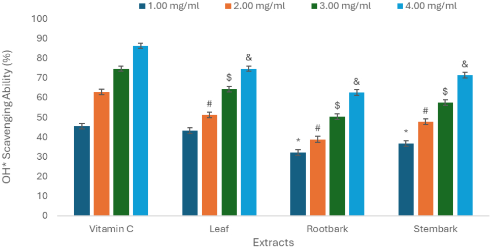
. Hydroxyl (OH*) radical-scavenging ability of aqueous extract of different parts of Boswellia dalzielli
Table 2 and Figure 5 display the extent to which the plant parts being studied can neutralize hydroxyl (OH*) radicals. The evaluation was conducted within the concentration range of 1.00–4.00 mg/mL. The leaf aqueous extract exhibited significantly more OH* radical-scavenging capacity (p < 0.05) compared to the other plant parts analyzed (Figure 5). Table 2 presents the SC50 values for three different plant sections. The leaf aqueous extract exhibited the lowest SC50 value (1.53 ± 0.02 mg/mL), followed by the rootbark and stem bark aqueous extracts with SC50 values of 1.94 ± 0.00 and 2.71 ± 0.03 mg/mL, respectively. The ability of vitamin C to scavenge OH* radicals at a concentration of 1.0 mg/mL was not significantly different (p < 0.05) from that of the leaf aqueous extract (Figure 5). However, all parts of the plant showed a significant OH* radical-scavenging capacity in a dose-dependent manner.
Figure 5
Hydroxyl (OH) radical-scavenging activity of aqueous extract of different parts of Boswellia dalzelli. Results are illustrated as mean ± S.D from 3 measurements. Results are significant at *p < 0.05 compared with vitamin C at 1.00 mg/ml; #p < 0.05 compared with vitamin C at 2.00 mg/ml; $p < 0.05 compared with vitamin C at 3.00 mg/ml; &p < 0.05 compared with vitamin C at 4.00 mg/ml.
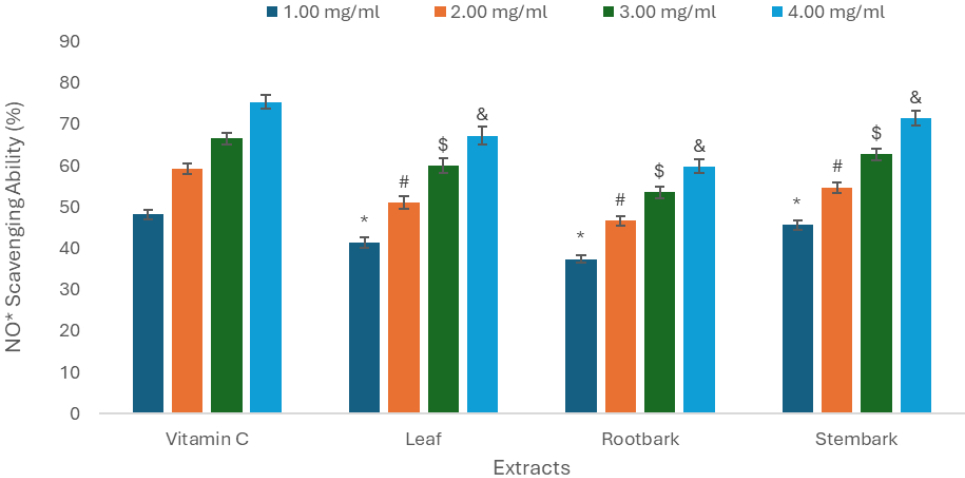
. Nitric oxide (NO*) radical-scavenging ability of aqueous extract of different parts of Boswellia dalzielli
The findings demonstrated that the plant components scavenged the NO* free radical in a manner that was dosage dependent (Figure 6). Based on the SC50 values reported in Table 2, it can be deduced that the plant components have varying levels of NO* free radical scavenging ability. Specifically, rootbark (SC50 = 2.32 ± 0.02 mg/mL) has the lowest level of scavenging ability, while stembark (SC50 = 1.37 ± 0.00 mg/mL) has the highest level (Table 2). It is noteworthy that although the B. dalzielii extracts exhibited notable NO* scavenging activities, their efficacy was not as high as that of vitamin C, a widely recognized powerful antioxidant.
Figure 6
Nitric oxide (NO) radical-scavenging activity of aqueous extract of different parts of Boswellia. Results are illustrated as mean ± S.D from 3 measurements. Results are significant at *p < 0.05 compared with vitamin C at 1.00 mg/ml; #p < 0.05 compared with vitamin C at 2.00 mg/ml; $p < 0.05 compared with vitamin C at 3.00 mg/ml; &p < 0.05 compared with vitamin C at 4.00 mg/ml.
. UHPLC-DAD-MSn analysis
Figures 7 display the UV chromatograms of the plant’s constituents at 254 nm. The numbers represent chemical constituents according to retention time. The results and identification of the compounds are shown in Tables 3. The identification of the compounds depended on the analysis of UV–Vis spectra at the wavelength of maximum absorbance, in addition to the masses and fragmentations derived from the mass spectra. The obtained values were then compared with standard constituents or available literature. The phytochemical analysis identified several chemical compounds, including phenolic acids, flavonoids, and stilbenoids. Notably, stilbenoid was detected in all parts of the plant.
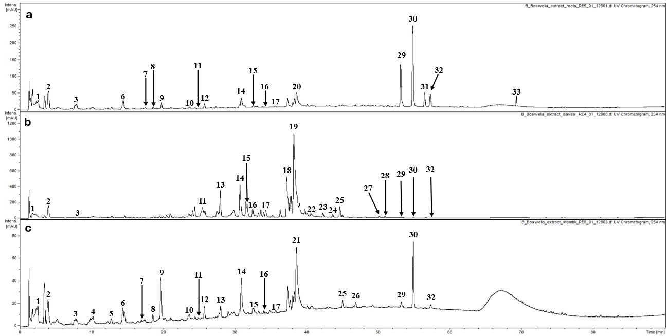
Table 3
UHPLC-DAD-MS data of constituents detected in the root, leaf and stem bark extracts of Boswellia dalzielli.
| No | Compound | Rt [min] | UV‑Vis [nm] | [M‑H]‑ m/z | MS2 ions | [M‑H]+ m/z | MS2 ions | Reference |
|---|---|---|---|---|---|---|---|---|
| 1 | Undefined compound | 2.6 | 278 | 355 | 337b, 319, 249, 193 | 357 | 339b, 321, 321, 293, 275, 265 | |
| 2 | Gallic acide | 4.1 | 269 | 169 | 125 | 171 | 153, 131, 127b, 109 | |
| 3 | Gallic acid derivativee | 7.8 | 270 | 343a | 325, 173, 169b, 125 | 345 | 327, 171, 153b | |
| 4 | Undefined compound | 10.2 | 270 | 305 | 179 | 307 | 289, 151, 139b | |
| 5 | Undefined compound | 12.8 | 274 | 593 | 575, 467, 441, 423b, 305, 285, 273 | 595 | 443, 427b, 409, 307, 301, 287, 283, 247, 229 | |
| 6 | Undefined compound | 14.4 | 271 | 495 | 477, 343b, 325, 193, 169 | 497 | 479b, 443, 327, 309, 263, 153 | |
| 7 | Undefined compound | 17.1 | 270 | 431* | 307b, 247, 163, 145, 125 | 433* | 415, 379, 361, 343, 317, 311, 309, 295, 287b, 271, 256, 233, 215, 191, 161, 149 | |
| 8 | Undefined compound | 18.6 | 274 | 305 | 287, 219b, 179, 164, 137, 125 | 307 | 289, 151, 139b, 121 | |
| 9 | Chlorogenic acide | 19.7 | 240, 300, 323 | 353 | 191b, 179 | 355 | 163b, 219 | |
| 10 | Undefined compound | 23.6 | 280 | 463 | 444, 417b, 293, 235, 181, 166, 161 | – | – | |
| 11 | Undefined compound | 25.3 | 271 | 461 | 415b, 269, 161 | – | – | |
| 12 | Undefined compound | 25.8 | 279 | 475 | 343, 233, 221, 209, 191, 176, 167b, 149 | 477 | 405, 369, 345, 331, 315b, 297, 279, 261, 243, 219, 207, 169 | |
| 13 | Ellagic acid derivativee | 27.9 | 266, 356 | 633a | 463, 301b, 329, 275, 257, 245, 229, 213 | 465 | 363, 321, 303b, 277, 259 | |
| 14 | Ellagic acid hexosidee | 30.9 | 252, 359 | 463 | 301 | 465 | 303b, 285, 131 | |
| 15 | Quercetin galloyl hexosidee | 31.8 | 254, 351 | 631 | 463b, 317, 179 | 633 | 615, 463, 319b | |
| 16 | Undefined compound | 32.6 | 273 | 437$ | 391b, 161 | 393 | 375 | |
| 17 | Methoxyquercetin hexosidee | 34.4 | 265, 353 | 479 | 317b, 179 | 481 | 462, 401, 385, 329, 319b, 296, 165, 153, 201 | |
| 18 | Ellagic acid pentosidee | 37.3 | 263, 352 | 433 | 301 | 435 | 303 | |
| 19 | Undefined compound | 38.0 | 259, 349 | 463 | 316b, 271 | 465 | 319b, 277, 137 | |
| 20 | Undefined compound | 38.6 | 250, 365 | 535$ | 447 | 537 | 449 | |
| 21 | Quercetin‑3‑O‑glucosidee | 38.9 | 265 | 509$ | 463, 311, 301b, 271, 190, 161 | – | – | |
| 22 | Undefined compound | 40.7 | 253, 354 | 447 | 432, 314b, 285 | 449 | 317 | |
| 23 | Undefined compound | 42.4 | 258, 352 | 433 | 343, 301b, 179 | 435 | 303 | |
| 24 | Undefined compound | 43.8 | 264, 350 | 599& | 447b, 313, 301, 285, 255, 179, 169 | – | – | |
| 25 | Astragaline | 44.8 | 255, 344 | 447 | 301b, 151 | 449 | 303b | |
| 26 | Undefined compound | 47.0 | 273 | 461 | 315 | 463 | 317 | |
| 27 | Undefined compound | 50.3 | 265, 350 | 431* | 285 | 433* | 287 | |
| 28 | Undefined compound | 51.1 | 219 | 551 | 483, 343b, 328 | – | – | |
| 29 | Methoxyresveratrol derivative | 53.4 | 317 | 741$ | 695b | 697 | 557, 473, 390, 383, 347, 333, 291b, 287, 279, 249 | Atta-ur-Rahman et al., 2005 |
| 30 | Methoxyresveratrol derivative | 55.0 | 316 | 549 | 307, 241b, 226, 163 | 551 | 389b, 293, 267, 255, 189 | Atta-ur-Rahman et al., 2005 |
| 31 | Methoxyresveratrol derivative | 56.6 | 317 | 595$ | 549b | 551 | 405b, 389, 375, 279, 185 | Atta-ur-Rahman et al., 2005 |
| 32 | Methoxyresveratrol derivative | 57.4 | 316 | 449$ | 403b, 265, 241, 226, 161 | 405 | 341, 267, 243b, 177, 135 | Atta-ur-Rahman et al., 2005 |
| 33 | Undefined compound | 69.4 | 219 | 241 | 225, 197 | 243 | 211, 135, 107, 91b |
. Discussion
The investigation of natural antioxidants has accelerated recently because of their possible health advantages and involvement in preventing disorders linked to oxidative stress. One plant known for its traditional therapeutic properties, B. dalzielii Hutch, has become a contender for these kinds of studies. This research provides a thorough analysis of the phytochemical composition and antioxidant capacity of aqueous extracts derived from the leaf, stem bark, and root bark of the B. dalzielii plant. This work aims to elucidate the diverse antioxidant capacities and identify the specific phytochemicals responsible for these effects using UHPLC-MS-DAD (Ultra-High-Performance Liquid Chromatography-Mass Spectrometry-Diode Array Detector) and in vitro tests.
Phenolic compounds can shield the body from free radicals, a common byproduct of aerobic cellular metabolism. According to Olanlokun and Akomolafe (2013) and Amic et al. (2003), they are potent antioxidants that can suppress oxidases, Eliminate free radicals, bind metal catalysts, stimulate antioxidant enzymes, and reduce alpha-tocopherol radicals. Table 1’s results show clear differences in percentage yield and bioactive profiles across B. dalzielli’s various sections. In comparison to the stembark and root bark, the leaf extract produced the highest extractive value, indicating a higher concentration of soluble phytoconstituents. This phenomenon may be attributed to the increased moisture and phytochemical contents found in leaves, which are generally more metabolically active plant tissues (Ma et al., 2022).
The phenolic content of the leaf extract was the highest, while the stem bark and root bark had the lowest concentrations. Phenolic compounds, known for their antioxidant properties, are abundant in the leaf extract, suggesting potential applications in the treatment of oxidative stress conditions (Heckmann et al., 2024). The leaf extract’s elevated phenolic content implies that it possesses a greater antioxidant capacity, which may contribute to its medicinal benefits. The flavonoid content followed a similar pattern, with the leaf extract exhibiting the highest levels, followed by the stem and root barks. Flavonoids are critical in the prevention of numerous diseases and inflammation reduction (Al-Khayri et al., 2022; Maleki et al., 2019). The leaf extract’s high flavonoid concentration underscores its potential to treat inflammatory diseases and enhance overall health.
The leaf extract also had the highest vitamin C concentration, which contributes to its strong antioxidant profile. Vitamin C is a well-recognized antioxidant that aids in the neutralization of free radicals and strengthens the body’s defensive mechanisms against oxidative damage (Akbari et al., 2022). The leaf extract’s increased amounts of vitamin C enhance its effectiveness as a robust antioxidant source. Remarkably, the stem bark extract showed the best ferric-reducing antioxidant potential (FRAP) despite the leaf extract having the highest phenolic and flavonoid concentration. This implies that the stem bark’s greater reducing activity may be due to the presence of additional strong reducing agents or the synergistic action of its phytochemicals. The root bark showed the least ability to reduce ferric iron, which is consistent with its lower concentration of bioactive compounds, even though it had the lowest content of total phenolic, total flavonoid, and vitamin C. The ferric reducing antioxidant power consistent with the concentration-dependent ferric reducing trend of aqueous extracts of Boswellia serrata reported by Sharma et al. (2011). These parallels reinforce the antioxidant potential of Boswellia species, supporting our conclusion that various parts of B. dalzielii, especially the leaf and stem bark, serve as potent sources of natural antioxidants capable of neutralizing reactive species through multiple mechanisms.
Overall, the differences in phytochemical profiles and antioxidant activities across the various sections of B. dalzielli emphasize the need of selecting specific plant parts for targeted therapeutic uses. The leaf extract is notable for its high total phenolic, total flavonoid, and vitamin C content, making it an attractive choice for antioxidant therapy. The stem bark’s considerable reducing capacity implies its potential use in oxidative stress-related disorders, whereas root bark, with its lower bioactive component content, may have fewer benefits in antioxidant therapy. The results of the present study are consistent with previous findings on the Boswellia genus. Kohoude et al. (2017) reported significantly high levels of total phenolic and flavonoid contents in the methanolic leaf extract of B. dalzielii, reinforcing the leaf as a potent source of bioactive polyphenols. Similarly, Brezo-Borjan & Švarc-Gajić (2024) examined resin extracts of Boswellia serrata obtained through subcritical water extraction and observed that both total phenolic and flavonoid contents increased with rising extraction temperatures. Although these studies involve different extraction techniques, they collectively highlight the high phytochemical potential of Boswellia species and support the findings of the current study.
Heavy metals have essential roles in several biological processes, such as cell growth and reproduction, the generation of biomolecules, multiple enzymatic reactions, and immunological function. However, excessive consumption of heavy metals may be dangerous. High amounts of heavy metal consumption may lead to toxic accumulation in the human body, which can pose significant hazards to several organs, including the neurological, pulmonary, reproductive, and digestive systems (Gulcin & Alwasel, 2022; Huat et al., 2019). This situation induces lipid peroxidation in the plasma membrane, resulting in the production of reactive nitrogen species (RNS) and reactive oxygen species (ROS) (Gulcin & Alwasel, 2022).
Fenton and Haber-Weiss reactions can also happen when transition metals like iron (Fe) and copper (Cu) are present. These reactions create reactive oxygen species (ROS) like hydroxyl radicals (OH*) (Bursal & Gulcin, 2011; Gulcin et al., 2011; Gulcin et al., 2005). The Fenton reaction, discovered by Fenton in 1984, involves the generation of hydroxyl radicals (OH*) by the interaction of hydrogen peroxide (H2O2) with metal ions and O2. The Haber-Weiss reaction, on the other hand, uses ferrous ions to help make hydroxyl radicals (OH*) from superoxide radicals (O2•) and hydrogen peroxide (H2O2). Both reactions have been identified as the main sources of radicals and the underlying factors behind cellular harm (Bursal & Gulcin, 2011; Gulcin et al., 2011).
Naturally occurring phytoconstituents derived from various plants, such as leaves, roots, stem bark, fruits, herbs, spices, and vegetables, have gained interest as potential antioxidants because of their wide range of bioactive properties and relatively low toxicity compared to synthetic antioxidants. These phytoconstituents have the ability to capture and neutralize free radicals, thus protecting the body from their harmful effects. We looked at how well different plant part extracts could chelate metals and get rid of free radicals. We did this by testing how well they could chelate iron (II) and copper (II), as well as their ability to reduce cupric ion and their DPPH and NO* radical-scavenging abilities.
The current study found that the water-based extract of different parts of B. dalzielli chelated iron (II) in a concentration depended manner (Figure 1). The leaf aqueous extract exhibited the highest Fe2+ chelating ability, indicating that it is the most effective iron chelator among the plant parts examined. The root bark extract has a greater SC50 value than the leaf and stem bark extracts, indicating a lesser chelating effectiveness. The SC50 value for EDTA, a standard chelating agent, was much lower compared to the plant extracts, demonstrating EDTA’s greater chelating activity. However, the leaf extract’s efficacy, comparable to that of EDTA at specific concentrations, underscores its potential as a natural chelating agent. These results indicate that the leaf extract’s bioactive components have a higher affinity for binding iron (II) ions, which could be related to its increased phenolic and flavonoid contents (Agarwal et al., 2021), as shown in Table 2. The stem bark extract exhibits substantial chelating ability, but to a lesser degree than the leaf extract. In contrast, the root bark extract’s decreased chelating ability could be attributed to a lower concentration of these bioactive compounds (Agarwal et al., 2021). Overall, the findings suggest that B. dalzielli leaf and stem bark extracts have strong iron (II) chelating activity, which may help treat disorders associated with iron overload (Jomova & Valko, 2011; Crisponi & Remelli, 2008). This result is strongly supported by the findings of Mohamed et al. (2015), who reported that methanolic and ethyl acetate extracts of Boswellia carteri resin exhibited significant Fe2+ chelating ability in a concentration-dependent manner. Their study found that the methanolic extract was the most effective, achieving 75.1% chelation at 0.5 mg/mL, although still lower than the standard chelator EDTA (IC50 = 0.028 mg/mL). The slightly reduced efficacy compared to EDTA observed in both studies does not diminish their potential, instead it highlights the relevance of these plant-based extracts as safer and more biocompatible alternatives for managing iron overload-related conditions. These findings collectively emphasize the promising role of Boswellia species, particularly B. dalzielii, in natural antioxidant therapy through metal ion chelation.
The in vitro copper (II) chelating ability of aqueous extracts from several sections of B. dalzielli demonstrated exceptional metal chelating ability. At doses of 0.83, 1.67, and 2.50 mg/mL (p < 0.05), the leaf extract demonstrated the most powerful chelating ability, with no significant difference from the control, EDTA. This shows that the leaf extract has a high affinity for copper (II) ions, similar to that of EDTA, a standard chelating agent. The stem bark extract also exhibited significant chelating activity, although to a lesser extent than the leaf extract.
In contrast, root bark extract has the lowest chelating activity. The reduced capacity of rootbark extracts to chelate copper (II) ions could be attributed to a lower concentration of active phytochemicals responsible for chelation (Jain et al., 2024; Torres-Rêgo et al., 2022).
EDTA exhibited superior overall chelating activity, however, the leaf extract also performed relatively well, emphasizing its potential as a natural chelating agent. These findings indicate that the bioactive chemicals in B. dalzielli leaf extract are particularly effective at chelating copper (II) ions, which could be advantageous in circumstances of copper overload (Kohoude et al., 2017; Onobrudu, 2017). The leaf and stem bark extracts’ significant chelating activity demonstrates their promise as natural options for regulating metal ions toxicity.
A compound’s ability to reduce cupric ions (Cu2+) to cuprous ions (Cu+), thereby contributing to the redox balance of the cell, is assessed using the cupric reducing antioxidant capacity (CUPRAC) assay (Silvestrini et al., 2023; Munteanu & Apetrei, 2021). The results of the CUPRAC experiment, presented in Table 2 and Figure 3, provide valuable insights into the antioxidant potential of various B. dalzielii extracts. According to the findings, there is a rising order for antioxidant capacity, as determined by the capability to remove cupric ions: rootbark < stembark < leaf. The rootbark had the lowest antioxidant capacity, while the leaf extract had the highest, followed by the stembark.
A detailed analysis of the data reveals that the antioxidant activity of the extracts increases with concentration (Figure 3), although the overall activity remains lower than that of vitamin C. This trend highlights the potential of plant extracts to act as antioxidants, particularly at higher doses. These findings are further supported by the SC50 values, which indicate the concentration required to achieve 50% of maximal antioxidant activity. The root bark extract exhibited the highest SC50 value (1.35 ± 0.13 mg/mL), indicating significantly lower antioxidant capacity, whereas the leaf extract showed the lowest SC50 value (0.64 ± 0.05 mg/mL), reflecting its superior antioxidant potential.
In the current investigation, B. dalzielli leaf extract outperformed stem and root bark in terms of antioxidant capacity. This result is in line with the Boswellia serrata investigation by Eryaman et al. (2024), which found that the leaf extracts had the greatest CUPRAC values. According to our data, B. dalzielli’s root bark had the lowest antioxidant capacity of all the parts that were examined. This is in line with findings from Boswellia serrata, which showed that the root bark had lower antioxidant activity than the leaves and resins (Mohammadi et al., 2017). This shows that all Boswellia species share a similar pattern in the distribution of antioxidant properties within various plant sections. Furthermore, the SC50 values that we obtained for the leaf extract of B. dalzielli (0.64 ± 0.05 mg/mL) are in line with the results obtained for Boswellia serrata, whose leaf extracts similarly shown strong antioxidant properties with low SC50 values (Mohammadi et al., 2017). This result demonstrates the potential of Boswellia leaves as a powerful antioxidant source.
The antioxidant activity of plant extracts is often evaluated using the DPPH (2,2-diphenyl-1-picrylhydrazyl) free radical scavenging assay, a widely recognized method for assessing non-enzymatic antioxidant potential. In this assay, antioxidants donate hydrogen atoms or electrons to stabilize DPPH radicals, leading to a visible color change from deep violet to yellow. The extent of this color change is directly correlated with the extract’s radical scavenging capacity (Ghosh et al., 2019; Thaipong et al., 2006).
In the present study, all tested parts of B. dalzielii, including the leaves, stem bark, and root bark exhibited DPPH radical scavenging activity in a concentration-dependent manner. Among them, the aqueous extract of the stem bark demonstrated the strongest activity, with a significantly lower SC50 value (0.77 ± 0.02 mg/mL) compared to the other plant parts. While this activity was somewhat lower than that of vitamin C (SC50 = 0.62 ± 0.01 mg/mL), it still indicates a potent antioxidant capacity. In contrast, the root bark displayed a higher SC50 value (3.26 ± 0.01 mg/mL), suggesting reduced efficacy at scavenging free radicals. These differences are likely attributable to variations in the concentration and composition of bioactive phytochemicals such as phenolics and flavonoids, which are known contributors to antioxidant activity (Hassanpour & Doroudi, 2023). These chemicals are generally found in larger concentrations in stem bark and leaves than in root bark, which may account for the observed variations in antioxidant activity.
The results of the present study are consistent with the strong DPPH radical scavenging activity reported by Kohoude et al. (2017) for methanolic extracts of B. dalzielii. Similarly, our findings are consistent with earlier studies on other species within the Boswellia genus. Mothana et al. (2011) reported that essential oils derived from Boswellia dioscorides, B. elongata, and B. socotrana exhibited DPPH radical scavenging activity in a dose-dependent manner. However, the antioxidant effects observed in those species at 1 mg/mL were relatively weak compared to standard antioxidant, ascorbic acid. In contrast, the stem bark extract of B. dalzielii in the present study displayed notably stronger activity at similar concentrations, suggesting interspecies variability in antioxidant potential within the Boswellia genus. Further supporting this trend, Awaley et al. (2020) demonstrated that Boswellia serrata stem bark extracts possess considerable antioxidant properties, aligning with our observation that the stem bark of B. dalzielii contains bioactive constituents capable of effectively neutralizing free radicals. While the antioxidant activity of B. dalzielii extracts was generally lower than that of vitamin C, a clear dose-dependent effect was observed, indicating enhanced activity at higher concentrations. This suggests that although B. dalzielii constituents may be less effective at lower doses, they still exhibit substantial antioxidant potential at elevated concentrations.
The ability of the extracts to inhibit the degradation of deoxyribose by hydroxyl radicals in the reaction mixture indicates their capacity to scavenge hydroxyl radicals. Singh et al. (2012) discovered that several plants belonging to the Boswellia family have antioxidant capabilities. Singh et al. (2012) found a significant accumulation of antioxidant chemicals in the bark of Boswellia serrata. The findings of this study suggest that Boswellia dalzielii represents a valuable source of antioxidants for pharmaceutical formulations and therapeutic interventions aimed at combating oxidative stress and reducing the harmful effects of xenobiotics or administered chemicals. Understanding the diverse radical scavenging characteristics can readily establish the correlation between the endogenous and exogenous antioxidant systems. The link between bioavailability and how well herbal medicines made from plants work in the body is very important (Jain et al., 2023; Munteanu & Apetrei, 2021; Rajput et al., 2021; Xie et al., 2021; Cekic et al., 2013).
In a physiological pH range, sodium nitroprusside produces the nitric oxide (NO) radical. According to Ashokkumar et al. (2008), it is a highly reactive substance that could alter the structural and functional behavior of numerous cellular components. Figure 6 illustrates a clear dose-dependent response to the study’s findings on the nitric oxide (NO*) radical scavenging ability of various B. dalzielii parts. Given the significance that NO* radicals play in oxidative stress and related diseases, the rootbark, stembark, and leaf extracts all showed the ability to scavenge these radicals. On the other hand, the scavenging potency varied among the plant sections.
The order of nitric oxide radical scavenging activity was as follows: stembark > leaf > rootbark. The stem bark extract, with an SC50 value of 1.37 ± 0.00 mg/mL demonstrated the most effective nitric oxide (NO•) scavenging activity among the plant components, highlighting its notable capacity to neutralize NO• radicals. Conversely, the rootbark had the lowest efficiency, as seen by its notably larger SC50 value of 2.32 ± 0.02 mg/mL. This implies that, in comparison to stembark, the rootbark may have lower concentrations or less effective antioxidant compounds compared to the stembark. The antioxidant activity observed in the present study aligns closely with findings reported by Sharma et al. (2011), who investigated the free radical scavenging capacity of aqueous extracts of Boswellia serrata. Their results demonstrated a strong, concentration-dependent antioxidant effect across various radical species, including nitric oxide (80.9%) and hydroxyl (40.97%) radicals at 500 mg/mL. These parallels reinforce the antioxidant potential of Boswellia species, supporting our conclusion that various parts of B. dalzielii, especially the leaf and stem bark, serve as potent sources of natural antioxidants capable of neutralizing reactive species through multiple mechanisms. Nitric oxide radicals are generated in biological cells and are involved in the control of several physiological functions. When oxygenated, nitric oxide is an extremely unstable species. Via intermediates NO2, N2O4 and N3O4, it interacts with oxygen to produce stable products nitrate and nitrite. The extracts’ ability to compete with oxygen and its derivatives in the current investigation explains their inhibitory potential against NO• radical (Sainani et al., 1997).
The phytochemical analysis showed the presence of a total of twenty (20) constituents in the root bark, including four phenolic acids (2, 3, 9, 14), two flavonoids (15, 17), four stilbenoid (29, 30, 31, 32), and ten unidentified compounds (Table 3, Figure 7). Of these, ten (10) compounds were fully or partially identified. Similarly, in the leaf aqueous extract, twenty (20) constituents were detected consisting of three flavonoids (15, 17, 25), five phenolic acids (2, 3, 13, 14, 18), three stilbenoid (29, 30, 32), and nine unidentified compounds (others). Eleven of the detected constituents were fully or partially identified (Table 3, Figure 7). Twenty-three (23) constituents were detected in stem bark aqueous extract, consisting of four flavonoids (15, 17, 21, 25), five phenolic acids (2, 3, 9, 13, 14), three stilbenoid (29, 30, 32), and eleven unidentified compounds (others). Twelve of the detected constituents were fully or partially identified (Table 3, Figure 7). A previous study utilizing high-resolution fast atom bombardment mass spectrometry (HRFABMS) identified stilbenoids as one of the bioactive constituents in Boswellia papyrifera, another species of this genus (Atta-ur-Rahman et al., 2005). In that study, the UV spectrum of the stilbenoid exhibited absorption peaks at 320, 306, and 218 nm (Atta-ur-Rahman et al., 2005). These absorption characteristics closely resemble the UV spectrum absorptions observed for the stilbenoid detected in the raw extract of B. dalzielii.
. Conclusion
The research demonstrates that different parts of B. dalzielii exhibit dose-dependent free radical scavenging abilities. Among the plant parts assessed, the leaf extract displayed the highest antioxidant activity across most parameters, suggesting a higher concentration or greater efficacy of antioxidant compounds. The root bark extract also showed considerable scavenging activity, likely due to the effectiveness or specific composition of its bioactive constituents. The phytochemical analysis revealed several chemical compounds, including phenolic acids, flavonoids, and stilbenoids, with a stilbenoid detected in all parts of the plant, although some compounds remain undefined. These findings suggest that B. dalzielii could be a valuable natural source of antioxidants, particularly in addressing disorders linked to oxidative stress. Ongoing investigations in our laboratory aim to isolate, identify and quantify the dominant compounds present in the plant extracts and elucidate their structures. The detailed quantitative and biological data on the dominant compounds will be presented in a follow-up publication.
